For decades, scientists believed that when a cell divides, its genetic material, called the genome, temporarily loses its 3D organization. And it consequently becomes a “blank slate” before rebuilding itself.
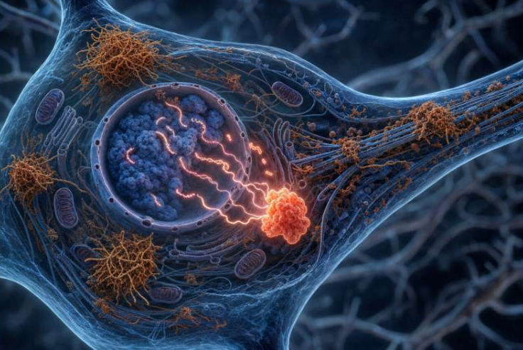
But a new study using a high-resolution genome mapping method has overturned that idea. Researchers have discovered that the genome actually retains an intricate 3D architecture during cell division, which are further supported by tiny, previously unseen structures called microcompartments.
This finding not only changes our understanding of how DNA stays organized during mitosis but also hints at a deeper link between genome structure and gene activity.
The Discovery and Methodology
The breakthrough came from a new method called Region-Capture Micro-C (RC-MC). It’s a way to map the genome with 100 to 1,000 times more detail than older methods like Hi-C.
To visualize how these changes unfold throughout the cell cycle, here’s a simple overview of microcompartment behavior at each stage of mitosis:
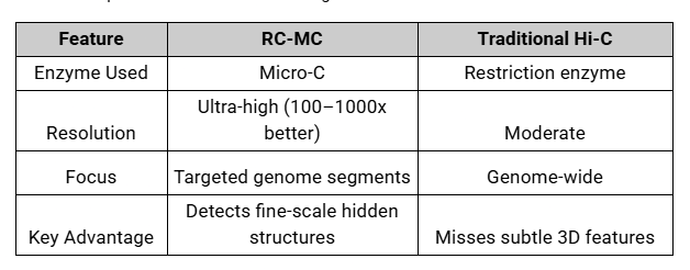
This new level of detail showed that the genome’s 3D structure never completely disappears. Instead, it stays organized by tiny, lasting links called microcompartments, even when most genes turn off during cell division.
“There is always structure — it never goes away,” the researchers noted, challenging the long-held view of mitosis as a structural reset.
What Are Microcompartments?
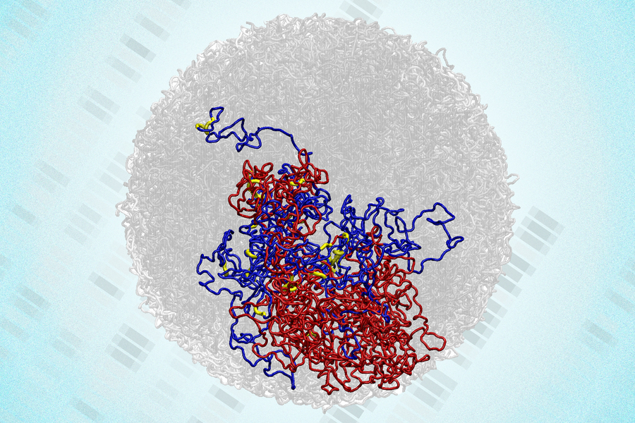
Microcompartments are tiny, highly connected loops of DNA formed when nearby regulatory elements, enhancers and promoters, stick together.
- Enhancers are DNA regions that boost the activity of genes, helping them turn on more efficiently.
- Promoters are DNA sequences where the gene-reading machinery binds to start transcription.
They represent a previously hidden level of genome organization, distinct from larger structures like A/B compartments or topologically associating domains (TADs).
Structure and Composition
- Definition: Small, densely connected loops linking enhancers and promoters.
- Formation: Created through an affinity-driven mechanism (similar to how block copolymers self-organize).
- Anchors: Mostly enhancer-promoter regions, unlike other loops anchored by CTCF proteins.
These loops act like “mini-hubs” for gene control, small neighborhoods where genetic elements interact closely to prepare for transcription.
Microcompartments During Cell Division
During mitosis, the cell’s genome undergoes dramatic changes. Larger structures such as TADs and A/B compartments vanish entirely. Yet, RC-MC revealed that microcompartments persist — and even strengthen.
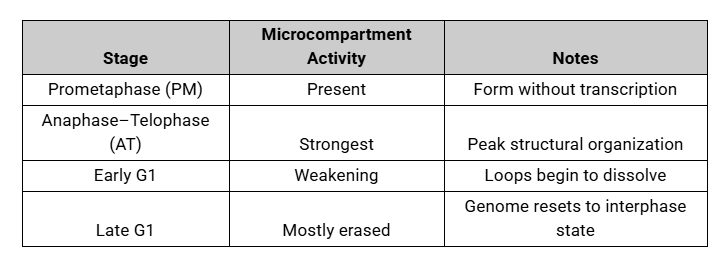
Surprisingly, microcompartments intensify during mitosis, especially in anaphase and telophase, when chromosomes are most compacted. The close proximity of enhancers and promoters appears to promote stronger looping interactions, helping preserve genetic “memory” through division.
The Physics Behind Microcompartments
To understand why these structures form and persist, the researchers ran polymer modeling simulations, computer-based experiments mimicking how DNA folds and interacts.
They identified three main physical drivers:
1. Chromatin Density and Compaction
- Higher density strengthens microcompartments.
- During mitosis, chromosomes are 1.5–3x denser than normal, creating an ideal environment for loop formation.
- Compaction reduces energy costs, favoring microcompartment stability.
2. Loop Extrusion Activity
- Loop extrusion proteins (like cohesin and condensin) can weaken microcompartments by disrupting local contacts.
- During mitosis, loop extrusion is reduced, which actually helps microcompartments stabilize.
3. Homotypic Affinity
- Regulatory elements with similar properties tend to cluster — a behavior known as homotypic affinity.
- This process resembles microphase separation, forming stable DNA neighborhoods even when transcription is off.
Linking Structure to Gene Activity
Interestingly, microcompartments don’t just persist, they appear to influence bursts of gene activity as cells exit mitosis.
Researchers noticed that genes with “transcriptional spikes”, meaning short bursts of activity, are located near stronger microcompartments. These genes form more loops, longer loops, and tighter connections during late mitosis.
This suggests that microcompartments help the genome “remember” which genes were active before division, a kind of molecular bookmark that guides the cell’s next cycle of gene expression.
Key Insights
- The genome’s 3D structure does not fully disappear during mitosis.
- Microcompartments persist and may play a memory-like role in reactivating genes.
- Physical forces, like compaction, affinity, and reduced loop extrusion, drive their formation.
- These structures may explain transcriptional spiking at the end of mitosis.
Takeaway
If the genome retains its fine-grained architecture even during cell division, perhaps DNA isn’t just a passive instruction book, as we all thought it to be, rather it’s an active, self-organizing system.
Microcompartments could be the hidden architects ensuring continuity between one cell generation and the next, isn’t it a fascinating idea into how resilient and cleverly arranged our biological code is? And there could be a possibility of hidden layers within our genome’s 3D world!
Frequently Asked Questions (FAQ)
Q1: What makes this discovery important?
A: It overturns the long-standing belief that 3D genome structure disappears during mitosis, showing that microcompartments preserve fine-scale genome organization even while larger structures vanish. This insight is crucial for understanding cell division and gene regulation.
Q2: What could this mean for biology and medicine?
A: Studying microcompartments may reveal how cells maintain gene activity through mitosis, which could impact gene therapy, cancer research, and cell-cycle biology. Understanding these structures might also guide innovations in genome engineering.
Q3: How do microcompartments differ from other genome structures?
A: Unlike larger structures such as TADs or A/B compartments, microcompartments are tiny, highly connected loops formed by enhancers and promoters. They persist during mitosis and may act as molecular bookmarks that guide transcription in the next cell cycle.
Q4: Why do microcompartments strengthen during cell division?
A: During chromosome compaction in mitosis, enhancers and promoters are brought into closer proximity. Combined with reduced loop extrusion activity from proteins like cohesin and condensin, this creates stronger and more stable microcompartment loops.
Q5: Could microcompartments affect gene activity in real time?
A: Yes. Microcompartments are associated with transcriptional spikes at the end of mitosis, indicating a direct structure-function relationship. They help certain genes activate quickly after division, ensuring continuity of gene regulation.