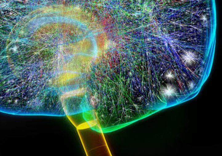
Cerebral cortex is the outer layer of cerebrum, that is the largest part of the brain. All kinds of higher-level processes such as language, memory, and decision-making are performed in this region.
The complex, densely packed structure is saturated with various types of neurons. It is extremely difficult to image its neuronal dynamics with the necessary spatial and temporal resolution.
So far, fMRI (functional magnetic resonance imaging) technique is widely used for studying brain activity. However, when it comes down to examine neural circuitry at the point of a single neuron, the result is poor temporal resolution.
Although it can capture small changes in blood flow associated with brain activity. But the technique has its limitations, as it cannot differentiate between:
- activities of different types of neurons, and
- distinguish between nearby regions of the cortex.
Wide field imaging technique to record cranial activity
Recently, neuroscientists started using wide field imaging technique to observe the cranial activity.
To carry out the research, neuroscientists at Duke University Medical Center and Cold Spring Harbor Laboratory measured activity of neurons in the cerebral cortex of mice using a series of genetic and imaging techniques.
This technique relies on genetically encoded sensors. These sensors can identify changes in the levels of calcium ions within individual neurons. Tracking calcium ions is an indicator of neuronal activity.
Distinct activation patterns of projection neurons
Josh Huang, one of the researchers who carried out the study explained that to study the activity of different types of neurons, they carried out the systematic approach.
They observed projection neurons – outside of the cortex to subcortical targets and to the thalamus – to gain insights into how different neural circuits are involved in behaviour and cognition.
Projection neurons are responsible for signal transmissions from one region of the brain to another. They are also crucial for learning, memory, movement, and perception.
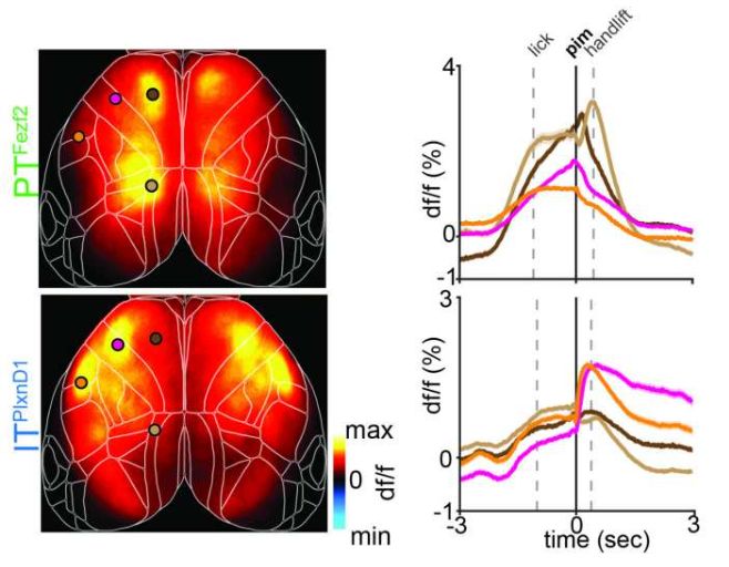
With the help of wide field imaging, the team was able to simultaneously monitor the activity of many neurons, across different regions of the brain.
It also enhanced their understanding of how the cortex is organized.

Variation in activity state of projection neurons
The neuroscientists concluded even though projection neurons are part of the same anatomical and functional units; they have distinct dynamic patterns of activity.
Generally, cortical neurons are arranged into columns that span multiple layers of the cortex and are considered both anatomical and functional units.
However, the research suggests that within these columns, different types of projection neurons exhibit unique patterns of activity that may be important for the function of the overall network.
Most importantly, Huang and colleagues found that the intratelencephalic (IT) and pyramidal tract (PT) populations of projection neurons (in the cortex) do not always function as a single unit. Even though they fall under the same anatomical and functional column.
Mode of operation
Researchers added that the IT and PT populations of projection neurons’ ‘mode of operation’ depends on the activity than an organism is doing at the present moment.
For instance, during a specific behaviour or task, the IT and PT populations may need to work together as a unit to coordinate and process information effectively.
Alternatively, during other behaviours or brain states, they may function independently, with the IT population contributing to certain aspects of neural processing while the PT population contributes to others.
Takeaway
This finding suggests that the functional organization of cortical circuits is more complex than previously thought.
Different types of neurons within a column may or may not act together. And this depends on the behavioural demands of the task involved.
This is a big step towards understanding many neurodegenerative diseases related to disruptions to specific neural circuits and cell types within the cortex.
The insight would surely assist in developing targeted interventions for neurological and psychiatric disorders.
Via: Medicalxpress


