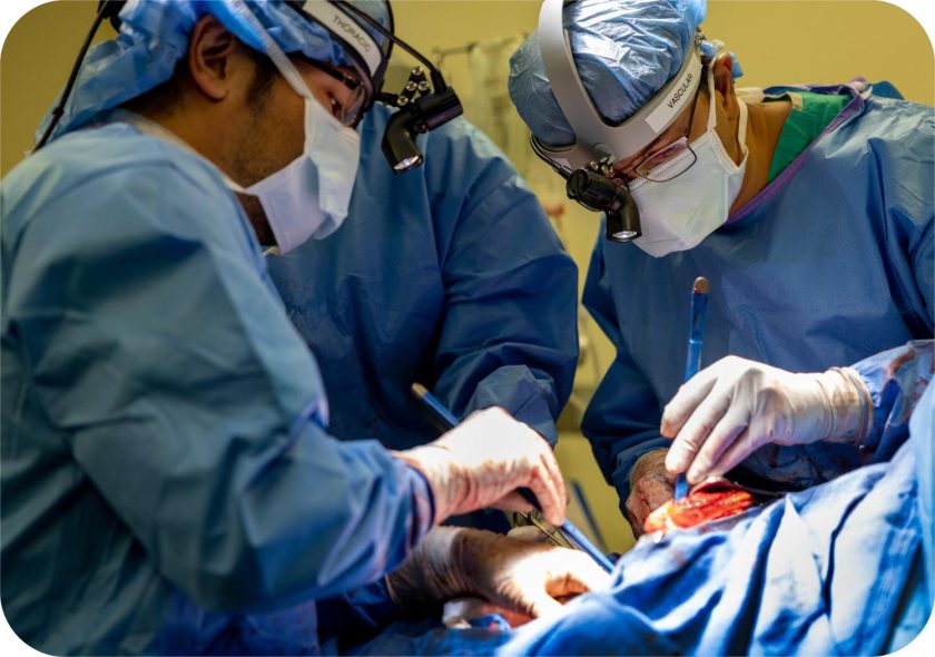
In recent years, 3D bio printing technology is being widely used by biotechnology firms and academia in tissue engineering applications with the help of inkjet techniques to create organs and other body parts. Using the innovative 3D bio printing technology, researchers have already created functional splints, valves and a human ear and are now trying to create a functional human heart for transplant, employing the patient’s cells. However, it would take years before one of these 3D printed hearts can be actually implanted in the human body.
A team of researchers from the University of Louisville, using cells for 3D printing, have achieved to print human heart valves and small veins and created other parts using some other techniques, said lead researcher and cell biologist Stuart Williams. The team has already tested the 3D printed blood vessels in mice and other small creatures. And in next 5 years, the team will be able to print parts to assemble a complete heart, says Williams. The printed heart so created using natural and artificial ingredients will be termed as bioficial heart.
According to researchers, the heart is a very complex organ and the biggest challenges while printing a heart is to make the cells used for printing, to work together as a team, as they do in an original human heart. On the other hand, the benefits using the cells from patients to print heart will solve the issue of rejection by the patient’s body that usually happens with artificial heart or with the donor’s heart. This would provide a fair chance of survival for patients who keep waiting for a perfectly matched donor, along with eliminating the requirement of anti rejection drugs, said Williams.
Although the technique uses cells from the fat tissue of the patients, there are still many challenges to overcome in creating complex organs as the heart and kidneys, such as to keep the printed tissue alive. Dr. Anthony Atala whose team of researchers is attempting to create a 3D printed human kidney at Wake Forest University, says that the team struggles to supply the structure with oxygen for survival, till the same gets transplanted and integrated in the body.
So if everything falls in place, then in less than a decade, the bioficial heart can be tested in humans, said Williams. Such bioficial hearts will be first implanted in patients with failing heart, who are not applicants for artificial hearts or for children that have a smaller chest to implant an artificial heart.
There are other techniques rather than 3D printing that are being used by the researchers to create a heart using cells from the patient’s body. Some other researchers are also using techniques in which the cells are put into a specific mold. In the laboratory, researchers have created a heart for rodents. Few simple body parts such as windpipes and bladders, have already been implanted in humans.
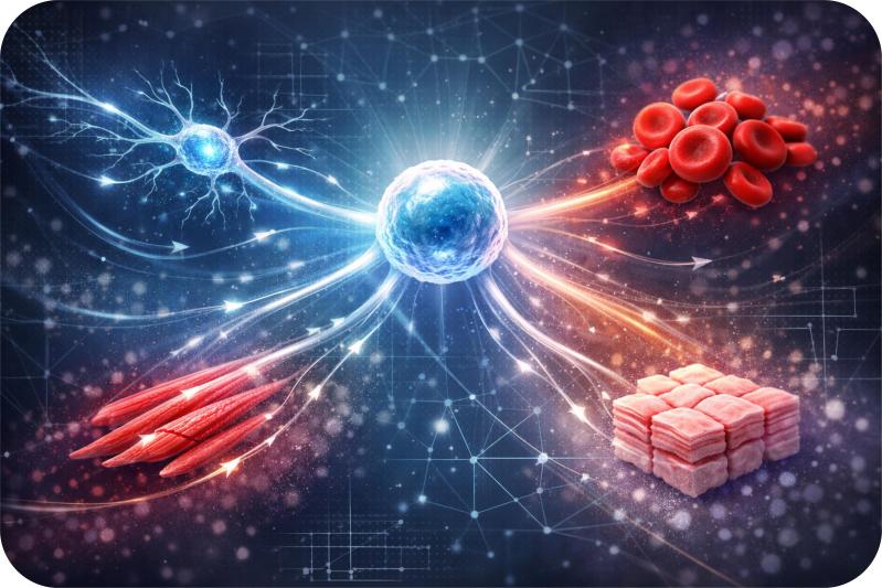
The 3D printers work similar to inkjet printers, employing needle that ejects material in a fixed arrangement. The cells used in 3D printing are purified in machines and using a computer model, printing begins to create the heart, layer by layer. The printer used by the William’s team uses a combination of living cells and gel to attain the desired shape of the organ, which gradually transform into tissues.
These techniques are also being employed in various other areas of medical science, such as building sure fitting prosthetics, a splint and an ear with living cells. The time is not far when the technology will be used for creating and implanting other bioficial organs for humans, saving lives of millions of patients waiting for a perfectly matched donor.
Source: Fox News


