Researchers at MIT have developed a wearable ultrasound patch that images organs without requiring an operator or gel.
The team has successfully demonstrated that the patch can, not only accurately images, but it can also gauge the fullness of the bladder.
This innovation has the potential to assist patients with bladder or kidney disorders. As it can monitor their organ function with greater convenience.
Wearable Tech Enables Early Detection of Deep-Seated Diseases
Researchers added that by adjusting the ultrasound array’s location and signal frequency, the device can also monitor various other organs. This versatility may facilitate earlier detection of deep-seated cancers like ovarian cancer.
Canan Dagdeviren, an MIT associate professor, describes the technology as a revolutionary tech that is capable of identifying and characterizing diseases in deep body tissues.
Diseases in deep body tissues often involve tissues that are not easily accessible for direct examination or diagnosis. For instance,
- Deep-seated Cancers: they affect deep tissues, such as ovarian cancer, pancreatic cancer, and certain types of lung cancer.
- Cardiovascular Diseases: conditions like coronary artery disease, heart failure, and certain congenital heart defects involve deep tissues within the heart and blood vessels.
- Kidney diseases: it includes polycystic kidney disease that affects the deep tissues of the kidneys.
- Neurological Disorders: conditions like brain tumors, deep brain injuries, or certain neurodegenerative diseases involve tissues deep within the brain and central nervous system.
- Respiratory Diseases: conditions like pulmonary fibrosis or lung cancer affect the deep tissues of the lungs.
Early detection and treatment of the above diseases are critical for effective medical intervention and improved patient outcomes.

Non-Invasive Ultrasound Patch for Internal Organs
Dagdeviren’s lab is known for creating flexible, wearable electronic devices. The team had innovatively crafted an ultrasound monitor intended for breast cancer screening. This device seamlessly incorporated into a bra.
Expanding on this expertise, the current team also applied a similar concept to develop a wearable patch. This patch adheres to the skin and is designed to capture ultrasound images of internal organs. Thus, offering a non-invasive means of imaging organs within the body.
The researchers, inspired by Dagdeviren’s brother’s experience with kidney cancer, chose to focus their first demonstration on the bladder.
After his kidney removal surgery, he faced challenges in fully emptying his bladder. This problem prompted Dagdeviren with the idea of a non-invasive ultrasound monitor, which could assess bladder fullness. And potentially aiding those with bladder or kidney issues.
Device Addresses Widespread Bladder Issues with At-Home Monitoring
With the device’s ability to monitor bladder volume, it can assist in the widespread problem of bladder dysfunction and related diseases, added Dagdeviren. As of now, measuring bladder volume involves traditional, bulky ultrasound probes in medical facilities. So, to solve this inconvenience, the team aims to create a wearable alternative for at-home use.
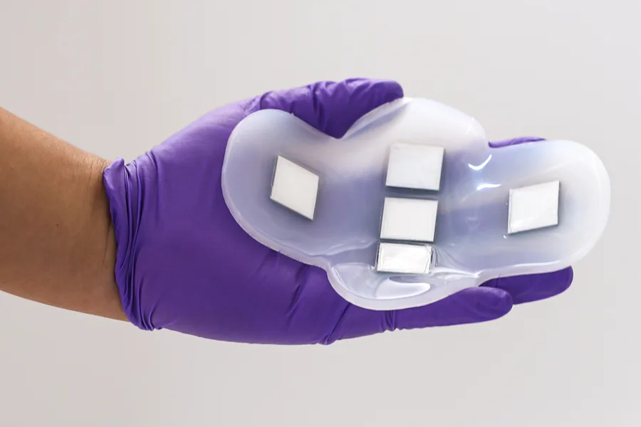
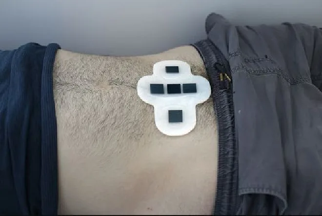
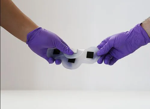
Their solution is a flexible patch made of silicone rubber. It features five ultrasound arrays crafted from a new piezoelectric material developed for this purpose.
Arranged in a cross shape, the arrays allow the patch to comprehensively image the bladder, which is approximately 12 by 8 cm when full.
The patch’s polymer is naturally sticky. This provides gentle adhesion to the skin for easy attachment and detachment. It can be comfortably secured in place with the help of underwear or leggings once applied to the skin.
Wearable Ultrasound Patch Matches Traditional Probes
In collaboration with the Center for Ultrasound Research and Translation and the Department of Radiology at Massachusetts General Hospital, researchers conducted a study. It demonstrated that their new patch can capture ultrasound images comparable to traditional probes.
Nearly 20 patients with various body mass indexes were involved in the study. Irrespective of the body mass index, the patch was able to give out high-quality images. It represented both – full, partially empty, and completely empty bladders, visibly.
Unlike regular ultrasound probes, this patch totally disregards the need for ultrasound gel and applied pressure.
Resultant images were viewed by connecting the ultrasound arrays to a standard ultrasound machine. Researchers envision to develop a portable device, roughly the size of a smartphone, for future image viewing.
Takeaway
Since, the tech is already a huge success, the MIT team is now actively pursuing the development of ultrasound devices for imaging various organs like the pancreas, liver, and ovaries.
For organs deep within the body, the team is exploring the possibility of the device functioning as an implant rather than a wearable patch.
The exciting results and vision pave way for future applications and the development of a portable device for image viewing. And potentially transforming the landscape of health monitoring with non-invasive and convenient solution.