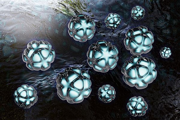
While working upon new security-related radiation detection, researchers at the University of Texas, Arlington discovered an advance in photodynamic cancer therapy. Wei Chen, professor of physics at the UT Arlington, noticed an odd luminescence emitted by copper-cysteamine (Cu-Cy) nanoparticles when while working on an experiment where he was exposing the nanoparticles to X-rays.
Upon further investigation, he found out that the luminescence was the byproduct of lost energy that the particles were diffusing. The same byproduct is also utilized in photodynamic cancer therapy to destroy cancer cells.
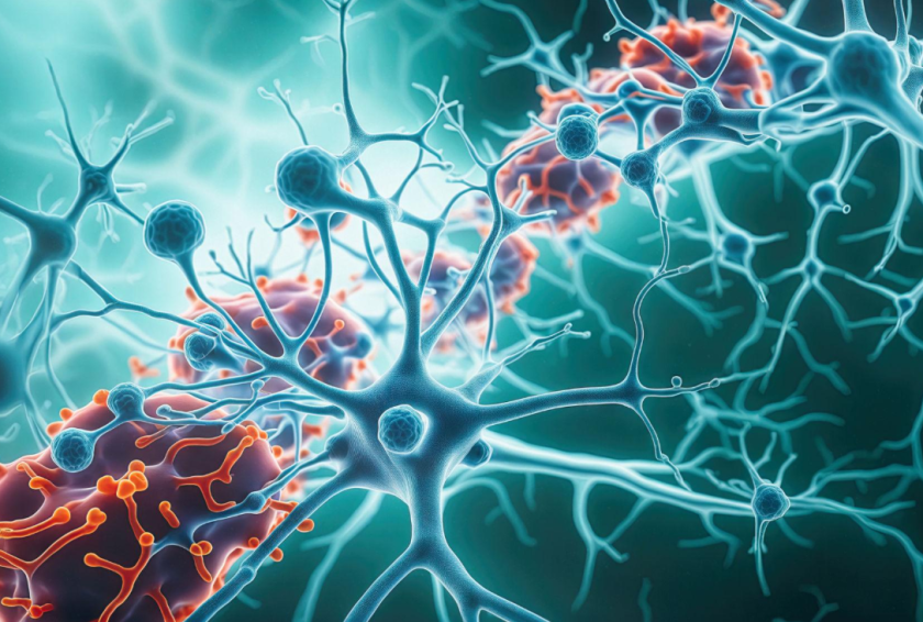
Photodynamic Therapy
Photodynamic therapy (PDT) is a method that is employed for providing damage to cancer cells. It involves introducing some photosensitizer into the tissue of tumor or the tumor site. Upon exposing light, the photosensitizer releases singlet oxygen – similarly as was experienced with copper-cysteamine (Cu-Cy) nanoparticles –that destroys the multiplying cells.
In 2012, researchers at Rice University tried the technique involving the gold nanoparticles, where they introduced nanobubbles using lasers at the tumor site. The act of bubbles bursting created small pores in the cell membranes, which then became a passage to deliver drugs.
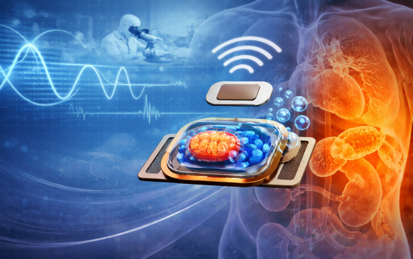
Combination of Cu-Cy Nanoparticles and X-Rays
Light penetration is one of the major huddles in PDT techniques, which makes the activation of nanoparticles a trickier business since the tumor resides deep into the tissues. However, with the discovery of Cu-Cy’s nanoparticle’s sensitivity to X- rays it seems that the problem could be solved.
X-rays’ ability of penetrating deep into the tissues of body allows for activating the photosensitizer with minimum effort and higher efficiency, plus the convenience is better than the previous photodynamic therapy methods.
Cu-Cy nanoparticle might also take another role, along with serving as photosensitizer it can well be used for cell imaging.
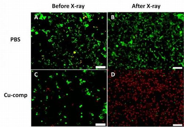
In order to prove its efficacy, Chen and his team conducted an experiment for more than 13 days. They employed the combination of Cu-Cy nanoparticle and X- ray over a tumor. After 13 days they noticed that the tumor under medicament did not grow in size but the tumor that was without treatment grew three times in size.
In order to be imbibed by tumor tissue, Chen and his team are working towards reducing its size. Currently, it’s around 250 nm, while the team is targeting below 200nm.
Once the desired size is accomplished, the innovation would be a breakthrough and this gives us a hope that one day we would be able to beat the multiplying cells effectively.
[…] In order to be imbibed by tumor tissue, Chen and his team are working towards reducing its size. Currently, it’s around 250 nm, while the team is targeting below 200nm. […]