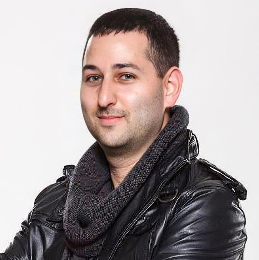
Dr. Maxwell Miner is a PhD candidate in the drug research doctoral program (DRDP) at the University of Turku working in the Roivainen group.
In the realm of scientific exploration, his focus lies in the intricate art of crafting preclinical radiopharmaceuticals, harmonizing their elements to illuminate the hidden nuances of inflammatory diseases and gliomas within the captivating world of animal models.
Moreover, he delves into the enigmatic realm of image analysis, channeling his expertise to refine the processing of colossal data sets and conjure visualizations that elevate our understanding to unprecedented heights.
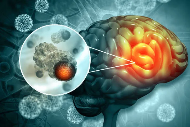
His recent work on “Targeting Folate Receptors in Gliomas to Detect Brain Tumors” caught our attention and so we tried to touch base with him for an interview. We got lucky, as he agreed to spend some time with us.
Dr. Miner here talks about how he has synthesized the imaging radiopharmaceuticals and built one of the semi-automated synthesis devices that they use routinely for projects. Scroll down to know more about his ideas and research 🙂
Would you please provide an overview of your doctoral research in the field of clinical physiology and isotope medicine?
Most of the projects I have taken the lead on revolve around PET imaging gliomas in experimental models. My hands-on duties are diverse and I always try to do as much as possible. For these types of studies, however, it is quite near impossible to do everything alone and I definitely rely on the rest of our group members to help out.
The work is so multidisciplinary, so it is great to pull from the expertise of scientists with differing backgrounds to help find unique solutions to whatever problems come up. And there are always a lot!
Nothing is quite straight forward in research and in this field, you often need to customize devices and figure out how to design an instrument to allow you to do an experiment and test some hypothesis. I deeply enjoy the problem-solving aspect of the work which sometimes requires a creative solution to the problem.
In addition to my own projects, I always try to help with other group members’ work when I can to “pay it back” or assist junior group members so they can really get their work moving forward, much like how past senior group members did for me.
Often, it is about helping troubleshoot issues they are having when things aren’t working as expected, performing some analyses, synthesizing some radiopharmaceutical, or showing how I would approach the data analysis. The Roivainen research group is a great multidisciplinary team and we work together super well.
In terms of hands-on work, I have synthesized the imaging radiopharmaceuticals and built one of the semi-automated synthesis devices that we use routinely for projects now.
These devices allow the radiochemist to make the compounds via remote-controlled valves behind a thick lead wall to lower their radiation burden. I have spent a lot of time also troubleshooting these systems and syntheses.
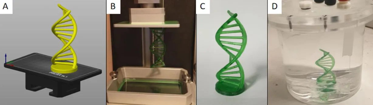
One hobby of mine is 3D-design and manufacturing which has allowed me to model and 3d print custom device parts, holders, and all sorts of things to help our work here at the Turku PET Center.
After these radiopharmaceuticals are made, they need to be rigorously tested for quality to ensure they are safe to inject. I have spent time developing and carrying out these analyses primarily via high performance liquid chromatography (HPLC), doing the same for metabolite studies where we want to assess what compounds they are breaking down into in the body (and at what rates). Data analysis is, of course, always a large portion of the work as well.
What motivated you to pursue these specific areas of study?
I sort of “fell into” it when searching for places to do a master’s project and felt that it all clicked together very well.
Earlier, I studied at the University of Ottawa in Canada and obtained a double major in chemistry and biochemistry. I also worked there on projects analyzing natural drug products or medicinal compounds in plants quantifying their amounts (via HPLC).
Later, I studied biomedical imaging in Turku Finland and reached out to multiple research groups at the Turku PET center here for a master’s project. After meeting with my now supervisors and starting to work here, I realized that it combined my background in chemistry, biology and biochemistry, and medical imaging all in one field- so it felt like the perfect match!
Previously I was feeling that all my studies were a bit disjointed and perhaps too much general knowledge without being exceptionally suited for any specific field, but radiochemistry really can utilize this diverse background exceptionally well. Overall, it feels that it just fits perfectly!
It is interesting to note that despite the city’s relatively small size (<200,000 in 2023), the Turku PET center is on the same playing field as the top universities in the world in this field of research. The history goes back to the early stages of the field and the leadership here has done a remarkable job over the years keeping things developing at such a rapid pace.
Globally speaking, such a top research and cancer imaging center is usually only found in some of the largest cities in the world with very advanced and large medical centers.
What are the primary applications of isotope medicine in both diagnostic and therapeutic contexts?
There are definitely wide and diverse applications with perhaps more even coming in the future.
Practically speaking, the scientific community has developed extremely sensitive devices to detect tiny amounts of radiation. If you are interested in a specific receptor or compound, you can attach a small radioactive nuclide to it or a compound that interacts with it and follow its accumulation or processing in the body in real-time.
For biochemistry and the study of living systems this is invaluable. The most common use, however, is for cancer imaging as it can provide a way to non-invasively biochemically distinguish between cells.
Can you share some examples of how radioactive isotopes are used in these applications?
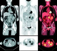
The most widely used radiopharmaceutical by far is fluorine-18-labelled deoxyglucose ([18F]FDG), which is an analogue of the same glucose sugar used for cellular energy.
It accumulates preferentially in cells which are using glucose at a higher rate. If you would see, for example, some “hot spots” or abnormal regions in a subject that have unusually higher [18F]FDG accumulation than you might expect, it is possible that it is a cancerous lesion that has increased energy needs due to it rapidly dividing. Thus, you can use this information to plan the treatment for the patient.
The PET-imaging field is much more than just cancer imaging though as you can attach a radionuclide to practically any interesting biological molecule, track where it goes and accumulates.
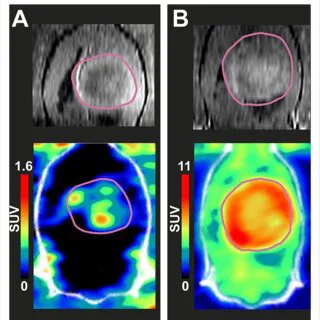
In the recent work we did with fluorine-18-labelled folate ([18F]Fol), we showed that there is significantly higher accumulation in glioma tissue than in the adjacent brain tissue in the experimental model.
There is a lot more to the field- from radiotherapy where the accumulation of the compound at a specific lesion provides radiation treatment therapy, or in sports science where radioactive oxygen can be inhaled while the subject does a muscle workout. The field has a lot going for it.
Radioactive oxygen can be inhaled and you can take a real-time video of the rate it accumulates in various muscles. There is a lot that can be studied.
Could you provide an example of a recent breakthrough in your field?
There are so many developments continuously coming forward. From new radiopharmaceuticals that target niche diseases such as prostate-specific membrane antigen (PSMA) for detecting prostate cancer, to improvements in PET camera technology. The field has so much more potential for growth.
In one of your research, “Exploiting Glutamine Consumption in Atherosclerotic Lesions by Positron Emission Tomography Tracer” I’m curious to know the significance of increased glutamine metabolism by macrophages in the development of atherosclerotic lesions?
In that work we proposed that “activated macrophages” start to consume glutamine and thus, absorb the radiolabeled 18F-glutamine much more than non-activated macrophages.
Macrophages are throughout most of the body waiting to fight some foreign invader or mitigate tissue damage and when they become activated send out chemical signals to “wake up” more macrophages and get them to accumulate at the same location to help fight whatever the problem is.
In that article we demonstrated that increased 18F-glutamine accumulation in these heart disease lesions is likely caused in part by activated macrophages and thus represents a possible way to detect and characterize the issue.
Simply put, if we see more signal from the heart in a PET image than normal, we can ultimately deduce that it is possible those regions may have ongoing heart disease.
What are your other interests besides academic commitments and research responsibilities… reading, painting, gardening, skiing maybe?
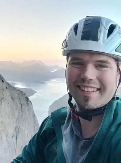
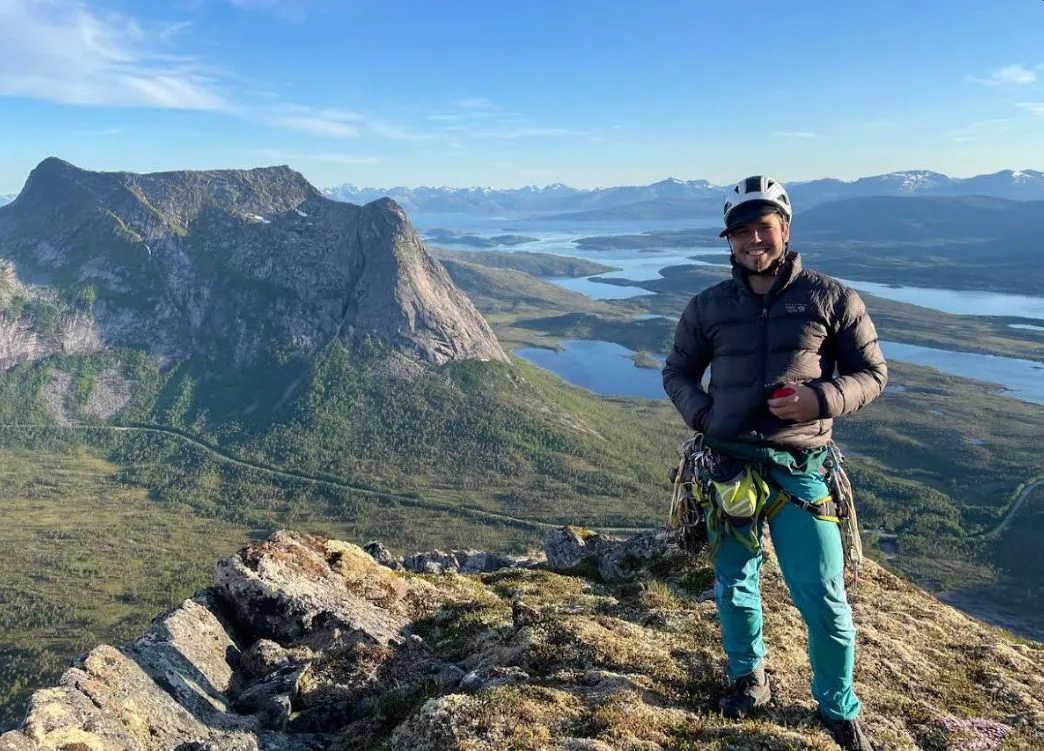
I love nature and outdoor sports in general, but I am completely obsessed with rock climbing and have been for quite many years.
I’ve worked part time in the climbing industry running courses, kids’ programs, rigging rope access work, and more. For climbing I really enjoy multi-day wall climbing which requires a lot of technical skill, rigging, and rope management.
It is all about problem solving in an extreme environment. Sleeping on a small portable ledge hanging off a wall really allows you access to some of the nicest camping sites in the world too. Most of the time you don’t have to worry about crowding! Hah!
Finally, what advice would you give to aspiring doctoral students interested in pursuing research in clinical physiology and isotope medicine? Are there any specific skills or qualities that you believe are essential for success in this field?
Just reach out to groups with your CV, apply, show some interest, have some relevant background skills, or have some scientific skills in general.
It’s amazing how diverse of a field it is- from robotics, 3D-design, physics and complicated mathematics, machine learning, imaging processing, chemistry, biochemistry, animal and experimental model development, tissue histology, and so much more.
It practically combines almost every other field of science in some way… perhaps barring astronomy- though when radiosynthesis fail randomly for unknown reasons you sometimes start to wonder if the planet alignment is influencing it in some way.
Quick bits:
What is your favourite movie quote?
From the Eiger Sanction:
You’re very good. I have really enjoyed climbing with you.
We’ll make it.
I don’t think so. But we shall continue with style.
If you were a superhero what would your powers be?
I’m not sure I’d want the responsibilities of having super powers. I would probably feel the need to dedicate my newfound abilities for the good of humanity which wouldn’t sit well with the current global power structure.
Your favourite scientific innovation of the 21st century?
Vertically landing rockets are pretty cool.
What will your TED Talk be 10 years from now?
Perhaps something about combining 3D printing with PET imaging camera calibration and development.
What books should I read in 2023?
I’ve recently been getting quite into history books revolving around large societal downfalls (e.g. roman empire) as well as social/societal revolutions (French, Russian, Haitian, British, American, and Spanish revolutions).
There are a lot of lessons to learn from the past so anything and everything in that space is exceptionally interesting. It’s amazing how impermanent things can be- from countries to thriving cities.
No one in Rome could have possibly guessed that the whole society would crumble. Much in the same way some giant company juggernaut of the recent past could have crumbled in changing times. Maybe we too, will live in interesting times. The wheel of history continues to roll forward at least!
Wow! Thank you, Dr. Miner, it has been a real pleasure! Your work is truly an inspiration. We look forward to visit you again and see more of your innovative research. Till then, we wish you all the very best for your future endeavor.